

July 2026
Dr. Eric Chow, OD
MIAMI — A child sits in a classroom that, to everyone else, looks perfectly ordinary. But to him, the hum of the air conditioner, the flicker of the lights, the shuffle of the student beside him, and the words on the page are all competing for attention at the same volume. He can't seem to sit still. He loses his place when he reads. By the end of the day he's exhausted, and no one can say why — his eye exam was perfect. He sees 20/20.
Down the hall in a different story, an adult six weeks out from a car accident describes the same kind of exhaustion. Grocery stores make her dizzy. Scrolling her phone makes her nauseous. Busy rooms feel overwhelming. Her scans are clean. Her vision, on paper, is fine.
Two very different people, and yet Dr. Eric Chow of Miami Vision Therapy hears versions of the same story every week. In both cases, the problem usually isn't how clearly the eyes see. It's a second visual system most people have never heard of — one that does its work long before you're ever consciously aware of it.
The main point: You don't have one visual system. You have two. One is slow, conscious, and built for recognizing and understanding what you look at. The other is fast, automatic, and built for orienting, protecting, and stabilizing you — before your conscious mind even gets a vote. When that second system is overwhelmed or underdeveloped, the result can look like inattention, clumsiness, sensory overload, or the lingering fog after a concussion.
To understand this, it helps to picture the brain in two broad territories.
The cortical system is the outer layer of the brain — the cortex, the wrinkled surface that includes the visual cortex, auditory cortex, motor and somatosensory cortex, and language areas like Broca's and Wernicke's. This is essentially the outer sheet covering all the lobes of the brain (frontal, parietal, temporal, occipital). It's where slow, conscious perception happens: recognizing a face, reading a word, understanding what you're looking at. It's powerful, but it's deliberate — and deliberate is slow.
The subcortical system sits underneath — the deeper, older machinery of the brain. This is the brainstem (midbrain, pons, and medulla), along with the thalamus, basal ganglia, cerebellum, and structures like the amygdala, hypothalamus, and hippocampus. These regions handle preconscious processing: primitive reflexes, posture, balance, threat detection, and the constant filtering of information you never consciously notice.
"Patients are surprised to learn how much of vision never reaches consciousness," says Dr. Chow. "Roughly ninety percent of the signal from the eye is routed toward the cortex for conscious perception. But an estimated ten percent branches off into these deeper, faster subcortical pathways — and that ten percent is doing an enormous amount of foundational work."
That foundational work includes orienting toward movement, detecting threats, triggering reflexive eye movements, gating attention, guiding posture and balance, controlling the pupil's response to light, and helping regulate your circadian rhythm. None of it feels like "seeing." All of it makes seeing — and functioning — possible.
Here's a striking way to see the difference between the two systems. Certain kinds of cortical injury can leave a person able to walk across a room, avoid furniture, and move confidently through space — yet unable to recognize the face of someone they've known for years. The fast, subcortical machinery that guides movement is intact; the slow, conscious machinery that identifies and understands has been disrupted. Two systems, two very different jobs.
Once you know both systems exist, a pattern that used to look like a coincidence starts to make sense.
Think about a child who can't sustain attention. Very often it isn't that the child won't focus — it's that the subcortical attention-gating system is overwhelmed. When the brain can't automatically filter out the background, everything floods in at once, and the child is left trying to consciously manage a firehose of sensory input. The same underlying immaturity often shows up as retained primitive reflexes — early-childhood reflexes that should have integrated and disappeared but instead keep interfering with posture, coordination, and attention.
Now think about a patient recovering from a traumatic brain injury or concussion. Why do so many of them develop balance problems, dizziness, vestibular symptoms, and sensory overwhelm in busy environments, even when their eyesight tests normally? Same answer: the subcortical system — the part responsible for orienting, stabilizing, and filtering — has been knocked off balance.
Key insight: Different populations, same underlying system. The developing child whose subcortical filters haven't fully matured and the injured adult whose subcortical filters have been disrupted can end up with remarkably similar symptoms: sensory overwhelm, poor balance, trouble sustaining attention, and fatigue. That's not a coincidence. It's the signature of a struggling subcortical visual system.
If there's one structure at the center of this story, it's the superior colliculus — a small hub sitting on the dorsal (upper) surface of the midbrain, on the roof of the brainstem. Its job is to coordinate where your eyes and head turn when something important happens in your environment.
It does this in two ways. Overt orienting is what it sounds like: actually moving your eyes and head to look directly at something — the fastest possible way to bring an object onto the center of your vision. Covert orienting is subtler: noticing something at the edge of your awareness without turning to look at it. It's how you sense someone approaching from the side, or track a movement in your periphery while keeping your eyes on the road.
"People sometimes assume covert orienting is just peripheral vision, but it's more than that," Dr. Chow explains. "Peripheral vision is where the information comes in. Covert orienting is your brain choosing to pay attention to it without moving your eyes. It's an attention skill, not just an anatomical one — and it's one we can assess and train."
One of the most elegant ways to check the health of this non-image-forming system is also one of the simplest: watching the pupil.
Most people know the basics — a pupil constricts when light hits it (the direct response), and the other pupil constricts too (the consensual response). But there's a more revealing test. A healthy pupil should stay constricted while a light is held steadily on it for roughly ten seconds. If the pupil starts to re-dilate sooner than that — while the light is still present — it can signal that the system holding it steady isn't in good control.
This is sometimes called watching for pupillary escape, or the "alpha-omega" pupil. Physiologically, the steady constriction is driven by the parasympathetic nervous system — your "rest and recover" mode. When the pupil escapes early, it often means sympathetic tone — your "fight or flight" mode — is overriding that calm parasympathetic response. In plain terms: a nervous system stuck in a heightened, defensive state can literally show up in how long a pupil can hold still under light. It's a small, objective window into whether the autonomic nervous system is in balance.
Even a single glance is the product of a remarkably orchestrated chain of events. Dr. Chow describes it in three legs.
First, the decision to look. The frontal eye fields (in the cortex) and the superior colliculus decide where and when to move the eyes. This is the command being issued.
Second, the routing. That command is sent to the correct gaze center. For horizontal movements, it goes to the PPRF (the paramedian pontine reticular formation, in the pons). For vertical movements, it goes to the riMLF in the midbrain. Think of these as the switchboards that translate "look over there" into precise instructions.
Third, the execution. The actual muscles are driven by three cranial nerves. Cranial nerve VI handles abduction (moving the eye outward). Cranial nerve IV handles intortion and depression (rotating the eye inward and helping it look down). Cranial nerve III does nearly everything else — elevation, most depression and adduction, raising the eyelid, and constricting the pupil.
Why this matters: An injury or weakness anywhere along that chain — the decision, the routing, or the execution — produces eye-movement problems. Which is exactly why careful assessment of how the eyes move, not just how clearly they see, can reveal where a breakdown is happening.
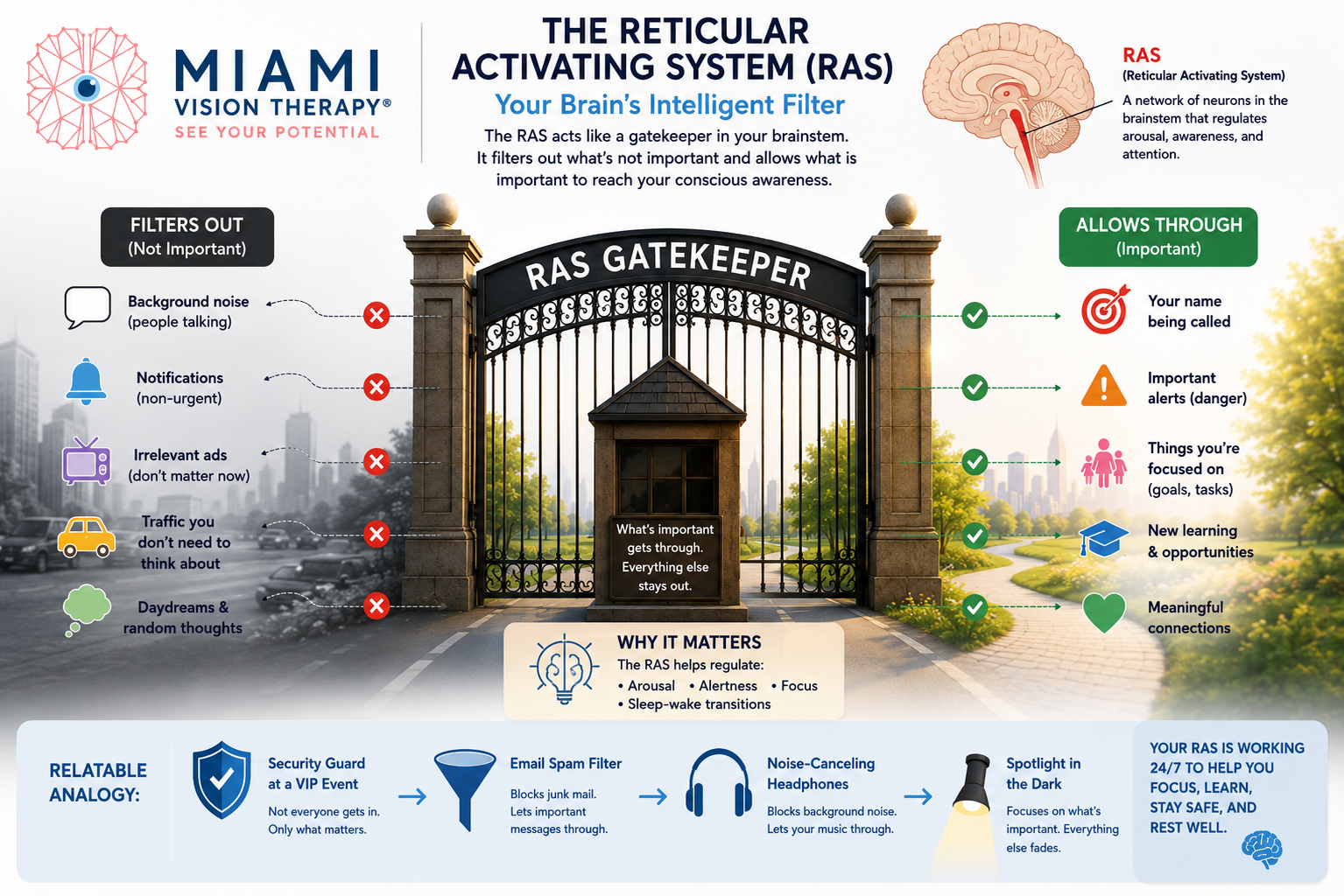
Underneath all of this sits perhaps the most important filter of all: the reticular activating system, or RAS. Dr. Chow likes to describe it as the gatekeeper of attention.
Its job is to decide what doesn't matter so that what does matter can reach your conscious awareness. Background noise, irrelevant ads, traffic that doesn't affect you, stray daydreams and random thoughts — the RAS filters these out so you can concentrate on what's in front of you.
The scale of that filtering is almost hard to believe. At any given moment, your senses take in an estimated 11 million bits of information per second. But the absolute most that can squeeze into conscious awareness is only about 40 to 50 bits per second. Everything else — the overwhelming majority of it — gets filtered out before you ever notice.
"When that gate isn't working well, the world gets loud," Dr. Chow says. "That's the child who can't tune out the classroom, and the concussion patient who can't tolerate the grocery store. Their conscious brain is being asked to manually process what a healthy filter should be handling automatically."
Key takeaways:
You have two visual systems: a slow, conscious cortical system for recognizing and understanding, and a fast, preconscious subcortical system for orienting, balancing, filtering, and protecting.
Though it carries only about 10% of the visual signal, the subcortical system does foundational work — attention gating, threat detection, reflexes, posture, and pupillary control.
When this system is immature (as in many children with attention challenges) or disrupted (as after a concussion or TBI), the result can be inattention, sensory overwhelm, poor balance, and fatigue — even with perfect 20/20 eyesight.
Structures like the superior colliculus, the eye-movement command chain, and the pupil's response to light give trained clinicians objective windows into how well this system is working.
Here's what gives Dr. Chow and the team at Miami Vision Therapy so much optimism: the subcortical visual system is not fixed. It's trainable. Through carefully designed vision therapy, it's possible to integrate retained reflexes, strengthen orienting and eye-movement control, calm an over-activated nervous system, and help the brain rebuild the automatic filtering it's supposed to do on its own. For children who struggle to focus and for adults working to recover after a brain injury, that can be the difference between white-knuckling through every day and finally feeling at ease in the world again.
If you or your child has been told that everything looks fine — but daily life tells a different story — it may be worth looking deeper than 20/20.
Schedule a functional vision evaluation with Dr. Eric Chow and the team at Miami Vision Therapy. Book online at miamivt.com/request-an-appointment or call (786) 558-7295.

Discover personalized eye care excellence with our comprehensive vision therapy services. Our expert team treats a wide range of vision conditions, including amblyopia (lazy eye), strabismus (eye misalignment), binocular vision disorders, double vision, post-concussion vision syndrome, and more.

For non-urgent questions or to learn more about our services, contact us today!
Managed and Designed by 